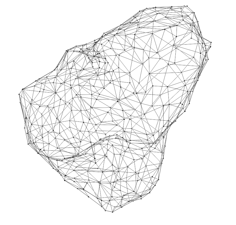
Hystological analisis through conventional 2D techniques has given enourmous help in understanding structure and function of different tissues. Nonetheless, the interpretation of cross-sectional plane sections skip significant information about the tisular architecture. Recent advances in microscopy and image analisis allowed the creation of 3D reconstructions with sub cellular resolution of tissues. Sadly, this is restricted to users with advance hardware and software.


Boris Vega, a member of the lab. In an effort to democratize the acces to this reconstructions, implemented an automatized work flux to optimize the geometrical models, thus, achieving their implementations in a web platform for visualisation. Boris’s tool is based in WebGL, which allows the user to visualisate, interact and extract information from the structures.
Click on the button to be redirected to the 3D visualisator.
